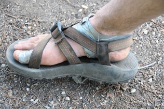
What Causes Blisters
 Today I found out what causes blisters.
We’ve all seen them, and unfortunately most of us have had one. Those painful, pockets of goo that form because some over exercised, over caffeinated, happy to be living life, outdoors-man decides to include you in their daily torture fest that is a hike up mount agony! So what exactly is going on here that causes the formation of a blister?
Let’s start off with the anatomy involved. For the most part, there are three layers of tissue that surround our bones and organs. The first layer is called the epidermis. This is the outer most layer of the skin. The second, or middle layer, is called the dermis. The third layer is called the subcutis, also known as the subcutaneous. Together, these three layers make up the largest organ in the body, our skin!
The epidermis itself has 4-5 layers. Most areas of the skin have four. In regions such as the fingertips, palms of your hands and soles of your feet, where the skin is exposed to greater friction forces, there are five. Starting from the deepest layer and working out, there is the Stratum Basale, the Stratum Spinosum, the Stratum Granulosum, the Stratum Lucidum and the Stratum Corneum. The Stratum Lucidum is that extra friction induced layer that helps make your skin thick in those areas that require it.
Friction blisters are just that, the result of friction. What’s going on under the surface is that repeated rubbing over one area of your skin will create forces that cause mechanical fatigue of the epidermis. This fatigue causes a split in the epidermis that usually resides at the layer of the Stratum Spinosum. When these cells separate, hydrostatic pressure allows a plasma-like blister fluid to form in this space. This fluid is similar to blood plasma in that it has the same electrolyte concentration, but with a much lower protein level. Should you continue the sadistic massage of your skin, the outer 3 layers of your puss pocket will break open, your salty fluid will leak out, and this portion of your epidermis will slough off. The resulting exposure to the last layer of the epidermis and the dermis itself (where your nerves are) is the red area that hurts like hell should you want to poke it with a stick!
The recovery of your exposed blister will begin after approximately 6 hours, as the remaining cells under your blister begin the process of healing. 24 hours into your rehabilitation, the cells are dividing like a mid-1990’s Microsoft stock. One day later, around 48 hours in to the restoration of your epidermis, a new Granular layer (Stratum Granulosum) can be seen. After approximately 5 days, this surge of cellular growth begins to slow down and a shiny new Stratum Corneum layer begins to show itself. Your new skin is ready!
Most blisters caused by friction do not require a doctor’s care. If the blister doesn’t pop, new skin will form underneath the affected area and the fluid is simply absorbed. Try not to puncture a blister unless it is large, painful, or likely to be further irritated. The fluid-filled blister keeps the underlying skin clean, which prevents infection and promotes healing. Should you need to burst your bubble, it might behoove you to follow a few simple techniques.
- Use a sterilized needle or razor blade (to sterilize it, put the point or edge in a flame until it is red hot, or rinse it in alcohol).
- Wash the area thoroughly, then make a small hole and gently squeeze out the clear fluid.
- If the fluid is white or yellow, the blister may be infected and needs medical attention.
- Do not remove the skin over a broken blister. The new skin underneath will benefit from this protective layer.
- Look for signs of infection to develop. These include pus drainage, red or warm skin surrounding the blister, or red streaks leading away from the blister.
Bonus Facts:
- Besides the common friction blisters, there are also blood blisters, burn blisters, chemical blisters, blisters caused by viruses and bacteria such as herpes and Ecthyma. Blisters from allergic reactions to things like poison ivy. There are also tiny fluid filled blisters, known as vesicles, that are associated with rashes. The difference between the many different types is the cause and appearance.
- A blood blister is a type of blister that forms when sub-dermal tissue and blood vessels are damaged without piercing the skin. It consists of a pool of blood and other bodily fluids trapped beneath the skin.
- Blisters bigger than 1.27 centimeters may be referred to as bullae.
- In most cases, the appearance of the blisters around the mouth or on the genitals, will allow a doctor to make the diagnosis of herpes. This diagnosis can be confirmed by a viral analysis of the watery fluid in a blister.
- Around 80 per cent of the adult population have antibodies in their blood, indicating past infection with HSV-1(oral herpes) and 25 per cent have antibodies against HSV-2 (genital herpes).
- Blister beetles are beetles (Coleoptera) of the family Meloidae, so called for their defensive secretion of a blistering agent, Cantharidin. There are approximately 7,500 known species worldwide.
- Cantharidin is toxic to people and animals. For centuries, cantharidin was prescribed as a cure for a variety of ailments. Spanishfly or cantharis, a preparation of dried meloid beetles, was thought to cure gout, carbuncles, rheumatism and many other medical disorders.
- The dried and crushed body of the blister beetle was once used medically as an irritant and diuretic, but was also regarded as a potent aphrodisiac, especially for older gentlemen before its dangerous nature was recognized.
- Today, the toxic properties of Cantharidin are more widely recognized and its use is largely restricted to veterinarians, who employ it as a counter-irritant and blistering agent.
| Share the Knowledge! |
|


